
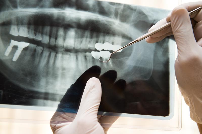
Dental imaging helps the dentist detect oral issues that aren't visible in a routine dental examination. It is required before any dental procedure and aids dentists in discovering hidden oral structures, benign or malignant masses, cavities, and bone loss.
Methods of Dental Image Processing
Traditional X-rays It is film-based dental imaging and helps to identify the disease within the skeletal structure. Dental images are made by directing an X-ray at the patient. A photograph film records the images that dentists then examine. Dental X-rays are used to identify:- Formation of periodontal pockets.
- The density of the bone around teeth.
- Prevalence of cavities, cracks, chips, fractures.
- Tumors in the dental cavity.
- Malocclusion.
- Abscess and cyst development in the bone and gum tissues.
- Incorrect positioning of teeth.
- Impacted teeth.
- Examining the development of permanent and baby teeth in kids.
- They cut the radiation levels by 70 percent.
- The dentist can view the photos on a computer screen.
- The photos can be enhanced, thus allowing the dentist to diagnose dental problems more efficiently and accurately. Dentists can detect tiny cracks that typically remain undetected by the conventional film.
- Diagnosis of sinusitis.
- Diagnosing patients suffering from gag reflex.
- Detection of abnormal growths in the mouth, neck, and head.
- Creating a customized treatment plan to enhance your appearance.
- Finding symptoms of oral cancer in the early stage.
- Assessing the progress of jaw disorders such as TMJ syndrome.
- Analyzing the signs of periodontal diseases such as gum and bone recession.
- Assuring proper growth of permanent teeth in kids.
- Making a treatment plan for braces, dentures, implants, and tooth extraction.